Classes

- Classes
We introduced online education in 2020. We know how difficult it is for you to get away from a busy practice, so we will be putting the didactic portion of our Soft Tissue Level 1 and Level 2 classes online. It is possible to purchase an 8 hour day of in-clinic, hands-on ultrasound training, on your machine, for an additional fee. This training will cover the scanning skills covered in the didactic modules, and will be RACE approved. We still offer regular Soft Tissue Level 1 and Level 2 classes in Irving, Texas, if you wish to join us here.
We have automated our online classes so that when you click on the link below, you then click on your class of choice, and will fill out a registration page, and put in credit card information for payment. If you opt in for emails, upon successful completion of a class, it will generate a CE. You will have access to PDF Notes and online Modules. Each class has 1 or more quizzes associated with it, that you must pass to get the RACE CE. If you do not pass, you can retake the quiz until you do. You will have six months to complete each class.
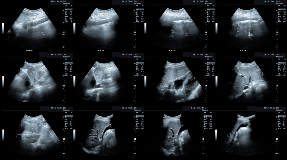
https://wave-we-are-veterinary-education.teachable.com/The objective for this course is for the attendee to leave with the knowledge of how to recognize an ultrasound case, incorporate ultrasound into their practice, and how to create a study for someone to interpret, of the five main abdominal organs. This will be accomplished through learning the basic knobs necessary to create a diagnostic image, how to tweak that image with these knobs, do measurements, and follow a scanning protocol. Recognizing artifacts will be covered, as well as how to quickly assess a trauma case with ultrasound. Taught by a Board Certified Radiologist.
Fee: Didactic Only 7 Hrs. CE: $300 per person, or $850 for 3 people.
Start Dates:
- Available anytime
Fee: Optional In-clinic hands-on 8 hour day, 7 Hrs. CE: $2100 with a Sonographer or $2400 with a DVM, in the continental United States. Call for a quote for Alaska, Hawaii, Canada, or overseas
Hands-on scanning will reinforce the didactic portion, and recognizing normal tissue.
The objective for this course is for the attendee to learn the normal and abnormal appearance of all abdominal organs, including pancreas, adrenal glands, and lymph nodes. In addition, scanning technique, machine controls necessary to create a high quality diagnostic image, measurements, and following a consistent scanning protocol will be emphasized. Ultrasound guided aspiration and biopsy technique will be covered. Taught by a Board Certified Veterinary Radiologist.
Fee: Didactic Only 10 Hrs. CE: $400 per person, or $1150 for 3 people.
Start Dates:
- Available anytime
Fee: Optional In-clinic hands-on 8 hour day, 8 Hrs. CE: $2100 with a Sonographer or $2400 with a DVM, in the continental United States. Call for a quote for Alaska, Hawaii, Canada, or overseas
Hands-on scanning will focus on scan technique, recognition of anatomy, and creating a diagnostic scan.
The objective for this course is for the attendee to leave with the knowledge of how to recognize an ultrasound case, incorporate ultrasound into their practice, and how to create a study for someone to interpret, of the five main abdominal organs. This will be accomplished through learning the basic knobs necessary to create a diagnostic image, how to tweak that image with these knobs, do measurements, and follow a scanning protocol. Recognizing artifacts will be covered, as well as how to quickly assess a trauma case with ultrasound. Hands-on scanning will reinforce the didactic portion, and recognizing normal tissue.
Fee: $1300.
Class Dates & Times: (Esaote sponsored means only Esaote equipment will be used)
- January 30 th , 2026 8 AM-5 PM
- March 25 th , 2026 8 AM-5 PM Esaote
- May 29 th , 2026 8 AM–5 PM
- July 30 th , 2026 8 AM-5 PM Esaote
- October 1 st , 2026 8 AM-5 PM
- December 10 th , 2026 8 AM-5 PM Esaote
The objectives for this class are for the student to learn knobs and buttons necessary to create an ultrasound study, how to tweak an image to be the best it can be, distance and area measurements, and following a scanning protocol for the liver, spleen, kidneys, and bladder. Discussing the purpose of each type of probe, and recognizing artifacts will also be covered.
FEE: $1100..
Class Dates & Times:
- January 30 th , 2026 8 AM-5 PM
- March 25 th , 2026 8 AM-5 PM
- May 29 th , 2026 8 AM–5 PM
- July 30 th , 2026 8 AM-5 PM
- October 1 st , 2026 8 AM-5 PM
- December 10 th , 2026 8 AM-5 PM
The objective for this course is for the attendee to learn the normal and abnormal appearance of all abdominal organs, including pancreas, adrenal glands, and lymph nodes. In addition, scanning technique, machine controls necessary to create a high quality diagnostic image, measurements, and following a consistent scanning protocol will be emphasized. Hands-on scanning will focus on scan technique, recognition of anatomy, and creating a diagnostic scan. Ultrasound guided aspiration and biopsy technique will be covered, and case studies will be presented. Taught by a Board Certified Veterinary Radiologist.
Fee: $1600.
Class Dates & Times: (Esaote sponsored means only Esaote equipment will be used)
- January 31st, 2026 8 AM-5 PM
- March 26 th , 2026 8 AM-5 PM Esaote
- May 30 th , 2026 8 AM-5 PM
- July 31 st , 2026 8 AM-5 PM Esaote
- October 2 nd , 2026 8 AM-5 PM
- December 11 th , 2026 8 AM-5 PM Esaote
The objectives for this class are for the student to learn equipment controls necessary to tweak ultrasound images to be their best, measurements, and following a scanning protocol to create a complete soft tissue ultrasound exam, which will be accomplished through hands-on scanning in a wet lab setting with a machine and animal.
Fee: $1200.
Class Dates & Times:
- January 31 st , 2026 8 AM-5 PM
- March 26 th , 2026 8 AM-5 PM
- May 30 th , 2026 8 AM-5 PM
- July 31 st , 2026 8 AM-5 PM
- October 2 nd , 2026 8 AM-5 PM
- December 11 th , 2026 8 AM-5 PM
The objective for this class is for the student to take their abdominal scanning skills to the next level. In depth discussion of hepatobiliary disease, vasculature evaluation with Doppler, and the search for shunts. The hands-on scanning will encompass:
- In depth evaluation of the liver, gall bladder, and bile duct, using standard subcostal and
intercostal windows
- Practice in finding and following the bile duct
- In depth evaluation of the canine and feline pancreas. Regional anatomy, including duodenum, stomach, transverse colon, portal vein, and splenic vein will be used to consistently find the pancreas or region of the pancreas
- Evaluation of the aorta, portal vein, caudal vena cava, renal arteries, hepatic artery will
be practiced, using Doppler (both color and pulsed wave) to assess waveforms, direction
and velocity of flow
- The portal vein will be evaluated closely for normal flow (direction and velocity) to better evaluate conditions such as portal hypertension
- Evaluation of portal vein and caudal vena cava for intra-hepatic and extra-hepatic portosystemic shunts
- Evaluation of the thyroid gland
Day 1: Lecture/Lab
Day 2: Live Cases
16 CE's
Fee: $1600.
Class Dates & Times: (Esaote sponsored means only Esaote equipment will be used)
- March 27 th , 2026 8 AM-5 PM Esaote
- March 28 th , 2026 8 AM-5 PM Esaote
- August 1 st , 2026 8 AM-5 PM Esaote
- August 2 nd , 2026 8 AM-5 PM Esaote
- December 12 th , 2026 8 AM-5 PM Esaote
- December 13 th , 2026 8 AM-5 PM Esaote
The objective for this course is for the attendee to leave with the knowledge of tweaking image quality, and learning both normal anatomy and appearance, as well as abnormal, and disease processes for the liver, spleen, pancreas, kidneys, adrenal glands, GI System, thyroid and vascular. Hands-on scanning will reinforce the didactic portion, and recognizing normal tissue.
Requirements: Must be a Boarded Specialist or Resident
Fee: $3900.
Class Dates & Times: (Esaote sponsored means only Esaote equipment will be used)
- October 5 th , 2026 8 AM-6 PM Esaote
- October 6 th , 2026 8 AM-6 PM Esaote
- October 7 th , 2026 8 AM-6 PM
- October 8 th , 2026 8 AM-6 PM
- October 9 th , 2026 8 AM-12 Noon
We will critique your imaging quality, protocol, and measurements, and help you to improve it.
10 cases for $650.
Cardiac Ultrasound
With each of these cardiac classes, we will offer a quantity of 10, Image Critique Packages, for an additional $750.00 per package, which allows for 10 ultrasound cases to be critiqued for image quality and selection, scanning technique, and assistance in creating a diagnostic scan. This does NOT include diagnosis of the case, but rather helping to create a diagnostic scan. Receiving constructive feedback on your cardiac scanning after attending a class, can help reinforce and tweak the skills you acquired in class.
We introduced online education in 2020. We know how difficult it is for you to get away from a busy practice, so we will be putting the didactic portion of some of our cardiac classes online. The optional hands on portion of these classes will be done in your clinic, on your machine. We will still offer cardiac classes in Irving, Texas, if you wish to join us here.
We have automated our online classes so that when you click on the link below, you then click on your class of choice, and will fill out a registration page, and put in credit card information for payment. If you opt in for emails, upon successful completion of a class, it will generate a CE. You will have access to PDF Notes and online Modules. Each class has 1 or more quizzes associated with it, that you must pass to get the RACE CE. If you do not pass, you can retake the quiz until you do. You will have six months to complete each class.
https://wave-we-are-veterinary-education.teachable.com/Canine MMVD
The new 2019 ACVIM MMVD Consensus statement guidelines recommends initiation of treatment with pimobendan in Stage B2 MMVD
The definition of Stage B2 was refined to reflect a specific heart size and diagnosis of Stage B2 MMVS is now the threshold for initiation of treatment with pimobendan in dogs with preclinical (Stage B) MMVD.
Echocardiography is the most sensitive method to measure heart size and is now the diagnostic test with highest priority in dogs with preclinical MMVD.
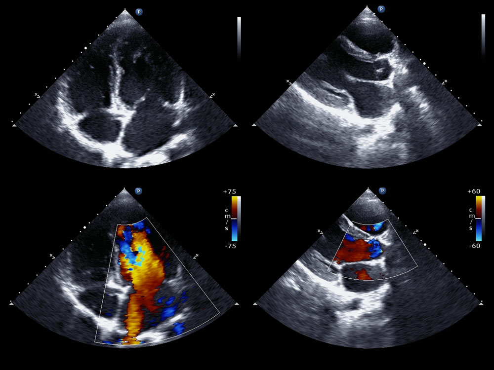
This course will teach you how to acquire and accurately measure the left atrium and left ventricle using simple right parasternal 2D echocardiographic images allowing you to diagnosis Stage B2 MMVD and recommend pimobendan treatment according to the 2019 ACVIM MMVD Consensus statement guidelines.
Feline Cardiomyopathy
The prevalence of feline cardiomyopathy is on average about 15% and increases with age
Many apparently healthy cats (20-50%) have heart murmurs, however many heart murmurs in cats are not pathologic.
Thirty percent of cats with preclinical hypertrophic cardiomyopathy go on to develop congestive heart failure (20%) and/or arterial thromboembolism (10%).
The best method to discriminate pathologic from non-pathologic murmurs is echocardiographic assessment with emphasis on accurate assessment of left atrial size
Left atrial enlargement represents a known risk factor for development of both congestive heart failure and arterial thromboembolism
In cats with active respiratory distress focused assessment of left atrial size can help support (left atria enlargement) or exclude (normal left atrial size) congestive heart failure
This course will teach you how to acquire and accurately measure the left atrium using simple right parasternal 2D echocardiographic images allowing you to stage the severity of cardiomyopathy.
This course is RACE approved and will require successful completion of quizzes covering the material and submission of 2 still frames and 2 videos that demonstrate your ability to acquire and correctly measure diagnostic images covered in this course.
The didactic portion of this course is available online and can be viewed at your convenience for 60 days after your registration date. There will be some live question and answer periods (up to 60 min) with the instructors, on dates to be determined. Attendees are encouraged to submit questions before (email) or during the live Q&A period. The Q&A period will also be recorded for those who are unable to attend live and be available for 2 weeks after the live event. There is one hands on scanning session planned for the center in Irving on October 10, 2020 for an additional fee as well.
CE: 7.5 Hours
Fee: $400. Per person, or $1150 for 3 people.
Fee: Optional In-clinic hands-on 8 hour day, 8 Hrs. CE: $2100 with a Sonographer or $2400 with a DVM, in the continental United States. Call for a quote for Alaska, Hawaii, Canada, or overseas
You can practice one on one instruction at your clinic, using your equipment, with up to 3 people.
Start Dates:
- Available now.
The objective of this course is to cover all right sided imaging planes - the core of an echocardiographic examination - as well as the left sided right auricular view. Subjective and objective evaluation of these images are covered. At completion of this course the attendee will be able to recognize all right sided imaging planes, understand the principles of obtaining these images, obtain critical measurements and understand their usefulness in the assessment of cardiac disease.
Fee: Didactic Only 6.5 Hrs. CE $350 per person, or $1000 for 3 people
Start Dates: Available anytime
Fee: Optional In-clinic hands-on 8 hour day, 8 Hrs. CE $2100 with a Sonographer, or $2400 with a DVM in the continental United States. Call for a quote for Alaska, Hawaii, Canada, or overseas.
Hands-on scanning will focus on image technique and knobs and buttons to create 2D right side and left cranial auricle views of the heart, as well as 2D and MMode measurements, to follow an imaging protocol.
The objective of this course is to cover two dimensional and left parasternal imaging planes in order to apply the principles of doppler as it applies to image quality and accuracy. Normal and abnormal color flow of MR, TR, RVOT, and LVOT will be taught in order to understand and incorporate spectral doppler and pressure gradients. Echocardiographic features of the common acquired diseases of CVD, PH, DCM, HCM, HOCM, and NSCM will be taught. At completion of this course the attendee will be able to incorporate color and spectral doppler into their echocardiographic exam, and recognize echocardiographic features of some of the common acquired heart diseases.
Didactic Only 11.5 Hrs. CE $550 per person, or $1600 for 3 people
Start Dates: Available Anytime
Fee: Optional In-clinic hands-on 8 hour day, 8 Hrs. CE $2100 with a Sonographer, or $2400 with a DVM in the continental United States. Call for a quote for Alaska, Hawaii, Canada, or overseas.
The objective of this program is to help the student understand the principals of Doppler and pressure gradients by examining the Spectral Doppler tracing and what affects it, what affects flow velocities, learn what normal systolic and diastolic pressures within the chambers & vessels of the heart are, and understanding and applying pressure gradient information.
Fee: Didactic Only 2 Hrs. CE $100 per person, or $275 for 3 people.
Start Dates: Available anytime
Cardiomyopathies are primary diseases of the myocardium characterized by a range of phenotypes that may or may not be progressive and may or may not result in clinical signs. Comorbid conditions such as systemic hypertension and hyperthyroidism can mimic feline cardiomyopathy and/or exacerbate disease progression if left unmanaged; therefore, these conditions should always be ruled out in cats with known or suspected cardiomyopathy across all stages (A–D). Initial clinical presentations may be acute and life - threatening, with common manifestations including congestive heart failure (CHF), arterial thromboembolism (ATE), and syncope.
This class is for Veterinarians and will emphasize practical, stage - based recommendations for the screening, diagnosis, staging, and treatment of feline cardiomyopathy, with a focus on hypertrophic and hypertrophic obstructive cardiomyopathy. The program will include hands - on echocardiographic imaging and measurement experience , with review of basic right - sided imaging planes used for assessment of left atrial size and function and for measurement of left ventricular wall thickness . A focused echocardiographic exam incorporating these elements is central to accurate phenotypic classification, risk stratification, and clinical decision - making. Key aspects of the ACVIM cardiomyopathy guidelines will be highlighted.
The lecture content will focus predominantly on the diagnosis, treatment, and potential prevention of CHF - related clinical signs—including dyspnea, tachypnea, weakness, and collapse —with emphasis on areas where new data have been published. Resources addressing current recommendations for the management of ATE are provided separately (CEG Cat ATE handout).
- Recognize and acquire basic right - sided (right parasternal) echocardiographic imaging planes commonly used in cats to assess cardiac structure and function.
- Evaluate left atrial (LA) size and function using appropriate echocardiographic techniques relevant to risk stratification in feline cardiomyopathy.
- Quantify left ventricular (LV) wall thickness using two - dimensional echocardiography and identify patterns consistent with hypertrophic cardiomyopathy (HCM) and hypertrophic obstructive cardiomyopathy (HOCM) .
- Interpret echocardiographic measurements of LA size/function and LV wall thickness to support diagnosis, phenotypic classification, and clinical decision - making .
- Apply findings from a focused echocardiographic examination to stage feline cardiomyopathy (ACVIM stages A–D) and guide screening, monitoring, and management.
- Describe the spectrum of feline cardiomyopathy phenotypes , recognizing variability in disease progression and clinical expression.
- Identify comorbid conditions —including systemic hypertension and hyperthyroidism— that may phenocopy or exacerbate feline cardiomyopathy and must be ruled out across all stages.
- Recognize acute and potentially life - threatening clinical presentations , including congestive heart failure (CHF), arterial thromboembolism (ATE), and syncope.
- Differentiate key diagnostic and management considerations between HCM and HOCM.
- Diagnose and manage CHF - related clinical signs in cats with cardiomyopathy, with emphasis on recently published data .
- Identify appropriate resources for evidence - based management of arterial thromboembolism (ATE) in cats.
Prerequisites: Cardiac Level 1, or ability to image right sided planes
2 Days: 12.5 CE’s
Fee: $2500
Class Dates & Times:
- August 12 th , 2026 9 AM - 6 PM
- August 13 th , 2026 8 AM - 4 PM
The rest of the course will cover:
The lecture content will focus predominantly on the diagnosis, treatment, and potential prevention of CHF - related clinical signs—including dyspnea, tachypnea, weakness, and collapse —with emphasis on areas where new data have been published. Resources addressing current recommendations for the management of ATE are provided separately (CEG Cat ATE handout).
- Features of Congenital Heart Disease
- For each condition the following objectives will be covered
- Background: prevalence, at risk breeds, characteristic murmurs, clinical signs, natural history, prognosis
- Salient 2D and M-mode features
- Salient color and spectral Doppler features
- Emphasis on diagnosis, staging of severity and prognosis
- Role of other diagnostic tests such as ECG and radiographs etc.
- Echo report considerations
- Referral considerations
10:00 AM – 3:00 PM
Lunch 12:00 – 1:00
10:00 – 12:00
Dr. Sonya Gordon and June Boon Introduction and Overview
Features of Congenital Heart Disease
- For each condition the following objectives will be covered
- Background: prevalence, at risk breeds, characteristic murmurs, clinical signs, natural history, prognosis
- Salient 2D and M-mode features
- Salient color and spectral Doppler features
- Emphasis on diagnosis, staging of severity and prognosis
- Role of other diagnostic tests such as ECG and radiographs etc.
- Echo report considerations
- Referral considerations
- Gradient review
- Hemodynamic information obtained from flow velocities
- Measuring spectral flow profiles
- Subaortic Stenosis
- Mitral and Tricuspid Dysplasia
- Ventricular Septal Defects
- PDA
- PS
- ASD
- Right to left shunts (reverse PDA, PFO, Tetralogy)
- Highlight use of contrast saline
10:00 AM – 3:00 PM
Lunch 12:00 – 1:00
10: 00- 12:00 June Boon
- Continue topics
- Subaortic Stenosis
- Mitral and Tricuspid Dysplasia
- Ventricular Septal Defects
- PDA
- PS
- ASD
- Right to left shunts (reverse PDA, PFO, Tetralogy)
- Highlight use of contrast saline
2 Days 8 CE’s Live Virtual
Fee: $1000
Class Dates & Times:
- October 17th, 2026 10 AM – 3 PM
- October 18th, 2026 10 AM – 3 PM
Cardiac Level 1: Diagnostic Two Dimensional Cardiac Scanning & Evaluation Accompanied by Common Cardiac Diseases
This course is designed to cover all right sided imaging planes - the core of an echocardiographic examination - as well as the left sided right auricular view. Subjective and objective evaluation of these images are covered. Hands on scanning sessions will teach the scanning technique needed to develop skill in obtaining these echocardiographic imaging planes. At completion of this course the attendee will be able to recognize all right sided imaging planes, understand the principles of obtaining these images, obtain critical measurements and understand their usefulness in the assessment of cardiac disease. This course will also include integration of echocardiography into your practice by covering the diagnosis and management of the most common adult-onset cardiac diseases. Using case material, we will form strategies for diagnosis, staging, and management of these diseases with emphasis on the role of echocardiography.
3 days ~ 21 hours of CE
Price includes 2 case critiques per week for 4 weeks after the class.
Fee: $2900
Class Dates & Times: (Esaote sponsored means only Esaote equipment will be used)
- April 18th, 2026 8 AM - 5 PM
- April 19th, 2026 8 AM - 5 PM
- April 20th, 2026 8 AM - 3 PM
- August 27 th , 2026 8 AM-5 PM Esaote
- August 28 th , 2026 8 AM-5 PM Esaote
- August 29 th , 2026 8 AM-3 PM Esaote
Cardiac Level 2: Doppler Echocardiography
The Complete echocardiogram: incorporating 2D imaging, measurements, and color and spectral Doppler.
The objective of this course is for the attendee to obtain an accurate and diagnostic echocardiographic study that incorporates color flow and spectral Doppler. All topics will focus on accuracy of the Doppler information and optimizing image quality. Topics covered will include: understanding the physical principles of Doppler echocardiography as it applies to the accuracy and quality of an echocardiographic exam, correct placement of Doppler cursors and gates, recognition of normal and abnormal color flow Doppler, understanding how Doppler aids in the evaluation of common acquired heart disease. Hands on sessions will focus on equipment controls that affect image quality, image optimization and measurement of the Doppler echocardiogram. Taught by a Boarded Cardiologist.
Prerequisites:/
Level 1 or experience obtaining right sided 2D and M-Mode images
2.5 days ~ 21 hours of CE
Fee: $2300
Class Dates & Times: (Esaote sponsored means only Esaote equipment will be used)
- June 1 st , 2026 8 AM-5 PM
- June 2 nd , 2026 8 AM-5 PM
- June 3 rd , 2026 8 AM-2 PM
- October 14th, 2026 8 AM - 5 PM
- October 15th, 2026 8 AM - 5 PM
- October 16th, 2026 8 AM - 2 PM
Cardiac Level 3: Advanced Doppler and 2D Echocardiography – Beyond the Basics
This course will address advanced Doppler techniques and diagnostics. At the conclusion of this course, the attendee should be able to:
- Evaluate diastolic function as it applies to cardiomyopathies in cats e.g. know how to assess functional diastolic class using pulmonary venous flow, mitral inflow profiles and isovolumic relaxation time
- Assess the risk of thrombus development in cats with cardiomyopathy by evaluation of left atrial function assessment using auricular flow and atrial fractional shortening
- Evaluate of left ventricular filling pressure and the risk for development of congestive heart failure in dogs using mitral inflow profiles, and isovolumic relaxation time
- Diagnose preclinical dilated cardiomyopathy in dogs: evaluation of systolic function beyond fractional shortening and systolic chamber size.
- Use echocardiographic methods to evaluate for the presence and severity of pulmonary hypertension when there is no tricuspid or pulmonary insufficiency.
Attendees should be able to obtain a basic 2D echocardiographic study including right and left sided imaging planes and have working knowledge and experience in color flow and spectral Doppler.
Prerequisites:
Cardiac Level 2 or clinical experience with Color and Spectral Doppler
2 days ~ 14 hours of CE
Fee: $1800
Class Dates & Times:
- December 3rd, 2026 8 AM - 6 PM
- December 4th, 2026 8 AM - 3 PM
Echocardiographic Features of Common Canine and Feline Acquired Diseases
The student will learn Two-dimensional, M-mode and Doppler echocardiographic features of common acquired diseases will be reviewed with emphasis on the essential images and measurements to establish a diagnosis and the generation of an echocardiographic report. Standard measurements will be reviewed. Staging and ACVIM consensus statement guidelines where appropriate will be highlighted. The final portion of the course will utilize case studies to reinforce course content. Minimal time will be allotted to disease management. The following is a list of the diseases that will be covered in this course. Laboratory time will be used to work through a standard 2D, M-mode and Doppler examination in addition to addressing any individual attendee needs. One laboratory session will be a hands on measurement session.
Dogs- Myxomatous mitral valve disease (MMVD)
- Pulmonary hypertension (PH) Dilated cardiomyopathy (DCM)
- Endocarditis
- Pericardial effusion & masses
- Cardiomyopathies (CM)
- Hypertrophic CM (HCM) / Hypertrophic obstructive CM (HOCM)
- Other CMs
- Non-specific CM (NSCM), Dilated CM (DCM), Arrhythmogenic right ventricular CM (ARVC)
- Transient myocardial thickening (TMT)
Upon completion the student will be able to scan, measure, and stage common canine and feline acquired diseases. This is a Medical class in a lecture/wetlab format.
Prerequisites:This course assumes attendees are familiar with right and left sided echocardiographic views and measurement and the utilization for spectral Doppler for assessment and interpretation of pressure gradients.
2.5 days 18 hours of CE
Fee: $2300
Class Dates & Times: ( Eacho Features of Common Canine and Feline Acquired Diseases )
- November 12 th , 2026 9 AM-5 PM Esaote
- November 13 th , 2026 9 AM-4:30 PM
- November 14 th , 2026 8 AM-2 PM
We will critique your imaging quality, protocol, and measurements, and help you to improve it
10 cases for $750.
We believe you learn best, if held accountable. This can be accomplished through a combination of constructive critical appraisal of attendee submitted imaging studies and outcome assessment. Outcome assessment will include practical laboratory examinations. Constructive critical appraisal of homework will provide attendees a basis to objectively assess their progress, recognize areas that require improvement and acquire the skills and techniques needed to become excellent scanners. Feedback of this nature in combination with outcome assessment is sure to escalate your personal needs with respect to becoming an excellent scanner.


The objective of this program is for the attendee to become a proficient, confident soft tissue sonographer, by immersing them in soft tissue ultrasound over a 3 month period. The student will be with us for 4 days in each session, with each session approximatel 1 month apart. Each session will build upon the prior session. Knobs, measurements, labeling, scan technique of the abdomen, scanning protocol, artifacts, normal and abnormals, disease processes, and aspirate and biopsy technique will be taught. Each session will require accountability in the way of quizzes, and written and practical exams. Case submissions will be required weekly between sessions for critique. Referral to a telemedicine service or specialist may still be necessary, as this is a skill set that requires continuing practice allowing proficiency to become excellence.
Requirements:
You may attend any one of these sessions as a "stand alone class", as long as you have the necessary clinical experience for each level. For example: no previous experience is necessary for Session 1, which is Beginner to Intermediate Level. Either Soft Tissue Level 1, Soft Tissue Immersion Program Session 1, or previous clinical experience would be necessary to attend Session 2, which is Intermediate to Advanced Level. Soft Tissue Level 2, Soft Tissue Immersion Program Session 2, or similar clinical experience would be necessary to attend Session 3, which is Advanced Level.
Attendees MUST have access to a digital ultrasound machine with color and spectral Doppler (PW required) capability, that allows saving and exporting still images and cine loops in Dicom format, for all three sessions.
Attendees will receive 24 hours of CE for each session, MUST attend all 3 sessions, and maintain a passing grade of 70 or above, per session, in order to receive the Certificate of Soft Tissue Proficiency. The scanning practicums and scanning homework will constitute the biggest percentage of the grade, as this class is geared to help you become an excellent scanner. Please do not schedule any vacations during this time, as not turning scanning homework in on time can seriously affect your grade. Please allow for up to 4 hours per week to do your homework. Doing homework on sedated animals will greatly benefit you, unless you have a couple of people that can assist in holding. The quality of your images is graded, and it is very difficult to get good images on a moving animal. This is NOT an accreditation. Class size is limited so we encourage you to sign up early to guarantee admittance. Taught by a Boarded Radiologist.
Attendees MUST have access to a digital ultrasound machine with color and spectral Doppler (PW required) capability, that allows saving and exporting still images and cine loops in Dicom format, for all three sessions. Attendees will receive 24 hours of CE for each session, MUST attend all 3 sessions, and maintain a passing grade of 70 or above, per session, in order to receive the Certificate of Soft Tissue Proficiency. This is NOT an accreditation.Class size is limited so we encourage you to sign up early to guarantee admittance. Taught by a Boarded Radiologist.
Fee per Session 1: $3900
Fee per Session 2: $3900
Fee per Session 3: $3900
Testimonials
Having tried other ultrasound courses, it wasn't until WAVE, that my
knowledge, skills & ability really exploded. Homework, in the Soft Tissue
Immersion Program, provides an opportunity to learn through repetition,
promoting muscle memory, while also receiving constructive criticism of
performance ability. WAVE is amongst the best veterinary CE in the world.
Cameron Dow, DVM
The Soft Tissue Immersion course provided the framework for a complete
abdominal ultrasound scan - far superior to any other ultrasound course I've
taken. I feel confident doing my own scans now.
Christie Macintyre, DVM
Class Dates & Times: (Esaote sponsored means only Esaote equipment will be used)
Session 1
:- April 8 th , 2026 8 AM-5 PM Esaote
- April 9 th , 2026 8 AM-5 PM
- April 10 th , 2026 8 AM-5 PM
- April 11 th , 2026 8 AM-3 PM
- September 22 nd , 2026 8 AM-5 PM Esaote
- September 23 rd , 2026 8 AM-5 PM
- September 24 th , 2026 8 AM-5 PM
- September 25 th , 2026 8 AM-3 PM
Session 2
:- May 13 th , 2026 8 AM–5 PM
- May 14 th , 2026 8 AM–5 PM Esaote
- May 15 th , 2026 8 AM–5 PM
- May 16 th , 2026 8 AM–4 PM
- October 27 th , 2026 8 AM-5 PM
- October 28 th , 2026 8 AM-5 PM Esaote
- October 29 th , 2026 8 AM-5 PM
- October 30 th , 2026 8 AM-3 PM
Session 3
:- June 17 th , 2026 8 AM–5 PM
- June 18 th , 2026 8 AM–5 PM Esaote
- June 19 th , 2026 8 AM–5 PM
- June 20 th , 2026 8 AM – 3 PM
- December 1 st , 2026 8 AM-5 PM Esaote
- December 2 nd , 2026 8 AM-5 PM
- December 3 rd , 2026 8 AM-3 PM
- December 4 th, 2026 8 AM-3 PM
This program is designed to meet the growing demand for veterinarians to become competent in the acquisition, measurement, interpretation, and reporting of diagnostic cardiac ultrasound exams in patients with acquired heart disease. Veterinarians will learn how to systematically interpret studies and prepare a comprehensive report. This program is also appropriate for Veterinary Technicians to develop the skills necessary to obtain and measure a complete echocardiographic examination appropriate for review by a supervising veterinarian or reading service.
Requirements:
- Must have access to a digital ultrasound machine with color and spectral Doppler capability
- Be able to save and export still images and cine loops in DICOM format
- Ideally a phased array transducer (cardiac probe) with CW capability is desirable but a microconvex is minimally required
This is a 4 session program, with approximately 8 to 14 weeks between each session (see dates below). Each week builds on the previous one. This course holds the attendee accountable for learning the didactic material and scanning technique in the form of daily quizzes, final written and practical exams as well as scanning homework between each of the onsite sessions. To successfully pass this course, participants must complete homework consisting of imaging studies on dogs and cats, and they must submit the images electronically in DICOM format for instructor review and feedback. The attendee can expect to spend about 2 hours per week on their homework studies. The homework is a mandatory requirement. If assigned echocardiographic exams are not completed each week, the attendee will not successfully finish this course.
Cardiac Immersion Program
Description
Session 1:
- Understand the practical physics of ultrasound.
- Identification of common echocardiographic artifacts.
- Learn patient positioning and preparation.
- Learn probe selection and equipment controls (optimize image quality).
- Learn relevant cardiac anatomy.
- Understand basic relevant cardiac physiology – sent as a pre-course assignment.
- Acquire all standard diagnostic two-dimensional cardiac right and left sided imaging planes
- Recognize normal cardiac ultrasound anatomy.
Description
Session 2:
- Review and optimize all right and left sided Imaging planes
- Learn how to obtain M-Mode images
- Learn and practice the common 2D and M-mode measurements
- Understand the principles of Doppler ultrasound as they apply to the cardiac color flow and spectral Doppler exam
- Acquire and identify normal and abnormal color flow.
- Color Doppler image optimization.
- Know the strengths and limitations of color Doppler.
- Acquire and identify normal and abnormal PW and CW Doppler
- Spectral Doppler image optimization
- Know the strengths and limitations of spectral Doppler
- Understand and apply the clinical utility of spectral Doppler with emphasis on the role of pulsed versus continuous wave
- Estimate pressure gradients and their clinical interpretation
Description
Session 3:
- Week 1 and 2 objectives will be reviewed and reinforced during week 3.
- Perform a complete 2D, M-Mode and Color flow Doppler ultrasound study including all standard measurements.
- Optimize and practice all imaging planes and the color flow and spectral Doppler obtained from these imaging planes
- Recognize the 2D, M-Mode, color and spectral Doppler features of common acquired canine and feline heart disease and the components of a comprehensive report for each disease
Description
Session 4:
- Week 1, 2 and 3 objectives will be reviewed and reinforced during week 4
- Using case studies:
- Reinforce the 2D, M-Mode, color and spectral Doppler features of common acquired canine and feline heart disease.
- Create a comprehensive report for each disease.
- Perform a complete 2D, M-Mode and color and spectral Doppler echocardiographic examination.
Who should attend?
- Veterinarians and veterinary technicians who wish to become excellent small animal imagers by being held accountable through written tests, practicums, and homework.
Prerequisites
- No previous scanning experience is required.
Fee per sessions 1, 3, 4: $3800 each
Fee per sessions 2: $4800 each
Class Dates & Times: (Esaote sponsored means only Esaote equipment will be used)
Session 1
- Small Animal Cardiac Immersion Session 1 will not be offered again until 2027.
Session 2
- Small Animal Cardiac Immersion Session 2 will not be offered again until 2027.
Session 3
- February 20th, 2026 8 AM-5 PM Esaote
- February 21st, 2026 8 AM-5 PM
- February 22nd, 2026 8 AM-5 PM
- February 23rd, 2026 8 AM-2 PM
- Small Animal Cardiac Immersion Session 3 will not be offered again until 2027.
Session 4
- April 13th, 2026 8 AM-5 PM Esaote
- April 14th, 2026 8 AM-5 PM
- April 15th, 2026 8 AM-5 PM
- Small Animal Cardiac Immersion Session 4 will not be offered again until 2027.
Click Here for a homework sample
We have automated our online classes so that when you click on the link below, you then click on your class of choice, and will fill out a registration page, and put in credit card information for payment. If you opt in for emails, upon successful completion of a class, it will generate a CE. You will have access to PDF Notes and online Modules. Each class has 1 or more quizzes associated with it, that you must pass to get the RACE CE. If you do not pass, you can retake the quiz until you do. You will have six months to complete each class.
https://wave-we-are-veterinary-education.teachable.com/Thoracic Radiographic Interpretation: A Review: 4.25 Hours of CE
This course is composed of a series of modules covering normal and abnormal interpretation of canine and feline thoracic radiology. Objectives of this course include review and assimilation of normal and abnormal aspects of the pulmonary parenchyma, mediastinum, pleural space, body wall, and heart. After completing this course, the participant will develop more confidence and expertise in interpretation of daily patient thoracic radiographs.
Fee: Didactic Only, 4.25 Hrs CE: $250 per person, or $700 for 3 people
Start Dates: Available anytime